Pages

Sunday, December 8, 2013

Good Bye Histology :(

My final week of Histology. I can't believe it. It's very saddening because it was a very enjoyable class.I'm really going to miss it.

As I reflect on this past semester, it's amazing how far I've come. When it first started, I was very scared of this class. I never knew what histology was and what it entailed. The first couple of weeks were hard. I had to learn how to adjust to the new teaching style and how to adjust my studying for this class. Eventually I got the hang of it. 

I brought my laptop to class look at the corresponding Powerpoints and take notes on them. Interaction was key for my understanding. I generally remained quiet unless I did not understand something. For exams, I figured that rewriting everything in a notebook along with highlighting key points and looking at pictures brought me a long way. With each exam, I got a higher score than before. I was learning and retaining the information.

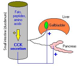
A lot of the information I was learning was overlapping with classes I took or was taking at the time. A lot of it was like Animal Physiology and some of it from Biochemistry. Honestly, I would have never done as well on my Biochemistry exams if it weren't for Histology. It was so awesome. This was the best semester I've ever had just because of Biochemistry and Histology. These two classes have really reinforced my love of science and learning. I have no doubt in my mind that I've exactly where I need to be. 

Sunday, November 24, 2013

Hugh Jackman and Basal Cell Carcinoma

Have you ever seen The Wolverine? Or any of the X-Men movies? How about Les Miserables? Hugh Jackman was in all of those movies and gave outstanding performances. I bet you're wondering why I'm talking about him. Well, I was driving to school one day when I heard that Hugh Jackman had basal cell carcinoma. That shocked me, and I felt sympathy for him.

But what is basal cell carcinoma? From what I've learned so far in histology, I was able to infer that it is cancer in the basal cells of the epidermis. That is just the general definition. From research, there can be a better explanation.

Basal cell carcinoma is when the basal cells in the stratum basal of the epidermis undergo uncontrollable growth. This can be caused by excessive exposure to UV rays. You see, melanocytes reside in this layer. Their function is to produce melanin to protect the skin from the UV rays. Luckily in carcinoma, the cancer isn't due to the melanocytes, so it is a nonmelanoma cancer. It is instead due to the uncontrolled growth of the tissue itself.

There are many forms of treatment such as medication, excision, electrodesiccation, cyrosurgery and many other options. He should be fine after he is treated. He needs to protect his skin from excessive UV ray exposure.


Reference: http://www.nlm.nih.gov/medlineplus/ency/article/000824.htm

Sunday, November 17, 2013

Digestive System Terms


Cholecystokinin - This is abbreviated as CCK. It is secreted from the mucosa of the duodenum. It's function is to stimulated the release of bile from the gallbladder and pancreatic enzymes. It also suppresses hunger.


Gastric Inhibitory Protein (GIP) - This protein induces insulin secretion from the pancreas. 


Somatostatin - This peptide is secreted in three places in the body: the stomach, the small intestine, and the pancreas. It reduces the secretion of HCl from the parietal cells in the stomach. It also prevents the release of gastrin, secretin, insulin and histamine.


Brunner's Glands - These are the glands found in the submucosa layer of the duodenum. It produces an alkaline secretion in the duodenum to protect it from the low pH of the chyme coming from the stomach along with neutralize it, activate the intestinal enzymes, and to lubricate the walls.

Saturday, November 9, 2013

Progressive Mulitfocal Leukoencephalopathy

As a biology major, the mechanism of this disease is so fascinating! Just thinking about it has my mind stuck in awe.

Progressive Multifocal Leukoencephalopathy is where the nerves in the brain become demyelinated, a disease caused by the JC virus. It is a rare disease. Even though it is a rare disease, this virus is found in about 85% of the adult population. How scary is that? The small silver lining in that is this virus remains dormant in the kidney, lymph nodes, and bone marrow (if you can call that a silver lining).

So then how does this virus cause this disease? Well, this virus is nicknamed the "opportunistic virus". This is because it waits until the body in under severe immunosuppression from HIV, drugs, or other diseases. When the body is at its weakest, this virus leaves its dormant state, travels to the brain, and destroys the myelination on the nerves by undergoing its lytic reproductive cycle.  This prevents signals from being sent to and from the brain. This can occur in many part of the brain, hence multifocal.  It is such a devastating disease with no cure or effective treatment.

------------------------------------------------------------------------------------------------------------

Falcó, V. (2013). Progressive multifocal leukoencephalopathy, a rare but devastating disease in AIDS patients. Indian Journal Of Medical Research, 138(1), 13-15.

Progressive multifocal leukoencephalopathy - a rare but serious disease. (2013). Australian Prescriber, 36(1), 24-25.

Wednesday, October 30, 2013

My Progress in Histology... and the Semester

I love love LOVE my histology class! It is such an engaging class. There have honestly been a few classes that I have been excited to go to; this class is one of them. By the grace of God, I have been improving my study habits and it shows in this class. To be honest, I did not think I would do as well I am now. Praise God!

This is such a relevant class. It feels like it is connecting a lot of the random knowledge I've been learning in college. It is also clarifying topics that I had trouble grasping. For example, it has helped me understand the nervous system so much better. In fact, I was assisting a professor with her class on Tuesday because she had trouble seeing. She was teaching her students about a part of the nervous system so she had be draw out pathways and terms on the board for her. It was so awesome because I understood everything she was saying! Histology has  really clarified and solidified so many concepts for me.

This is my second Reflection post. My final post will be a reflection on the class as a whole and its impact on my personal life and future career.

Saturday, October 26, 2013

Arteriosclerosis

Atherosclerosis is defined as paste and hardening in the arteries as athero means “paste” while scleroisis means “hardening”. Arteries are endothelium-lined blood vessels that transport oxygen- and nutrient-rich blood to the different organs and tissues of the body. These veins should be very flexible, sturdy, and elastic. As a person ages, they start to lose this elasticity and flexibility due to hardening of the endothelium and plaque accumulation, so the arteries stiffen and narrow to a degree which of course is inevitable. But this can also be influenced by many more factors aside from age such as diet, exercise, smoking, diabetes, high cholesterol, high blood pressure and presence of free radicals. This damages the endothelium and promotes further hardening and narrowing, which has adverse physiological effects in the body, particularly in the heart, brain, and peripheral regions like the legs.

The aforementioned factors that cause damage to the epithelium are by lifestyle and/or genetics. They cause an immune response that leads to an inflammatory response of plaque formation and impair the epithelium’s function of proper blood flow. This plaque is mainly composed of low-density lipoproteins (LDL) cholesterol because as the monocytes try to attack and engulf the cholesterol, they actually fuse and form larger cells called foam cells. These cells enlarge through mitosis and further accumulation of fat and cholesterol. This eventually hardens the epithelium and narrows the artery thus limiting the amount of blood that can flow through the artery. 
------------------------------------------------------------------------------------------------------------
Boamponsem, A.G., and L.K. Boamponsem. "The Role of Inflammation in Atherosclerosis." Advances in Applied Science Research 2.4 (2008): 194-207. Pelagia Research Library
Ulbricht, Catherine, ed. "Atherosclerosis: An Integrative Approach: A Natural Standard Monograph." Alternative and Complementary Theories 17.5 (2011): 287-93. Print.

Friday, October 18, 2013

Cardiologist Shadowing

I finally started my shadowing! Woo hoo!

My primary care doctor is both a general practitioner and a cardiologist. He mostly sees patients with referrals for stress tests or ECGs. It was pretty monotonous at times because he asked the same questions, and say a lot of the same things. One patient, though, sure made things interesting.

This patient had an ECG done that indicated he had suffered from a myocardial infarction. The machine detected it and so did the doctor. What was really odd is that the patient never experienced the pain of a heat attack. The doctor explained that it must have been a very minor heart attack. I was just standing there in awe of the situation. The patient then wanted to know what happened to the heart after the heart attack, so the doctor responded by saying that the heart forms scar tissue where the dead muscle cells are, making the heart less efficient.

All of this was just so cool to me. After seeing that patient, I told the doctor that I had just learned what happens when a person suffers a heart attack. When a person suffers a heart attack, some of the muscle cells die. In response, the fibroblasts in the heart muscle make collagen to fill in that area, which is the scar tissue formed on the heart. Since it isn't muscle tissue, it doesn't have any contractile abilities so it makes the heart less efficient in working as a whole and transmitting the electrical signal across the whole heart.

This encounter was so awesome!